Salem Operation Technique
General or Spinal anesthesia is used. The patient is supine. A 5 cm incision is made in the femoral skin crease with its lateral end over the femoral pulse. The saphenofemoral junction is exposed and all of its tributaries are divided and legated. The proximal par of the saphenous vein is transected between hemostats and the proximal stump is double ligated at the saphenofemoral junction.
A 2 cm transverse or longitudinal incision is made 1-2 cm above and anterior to the medial malleollous. The edges of the transected lower end of the saphenous trunk are grasped between two mosquito hemostats. The probe end of the stripper is advanced proximally through the entire length of the vein. At the femoral incision the cover of the probe is replaced by the head system, and the narrow ends of the heads are directed toward the wire.
The metal bar connections are used in tubular varices, whereas the wire connections are used in large, tortuous varices, and combinations of both are used according to the case.
The stripper is pulled downward gently inside the vein and any head larger than the vein must be removed. Then the upper end of the vein is ligated with a double ligature and the femoral incision is closed. As the stripper is pulled downward, the spikes of the heads abrade and scrub the intima into small fragments and sweep down the free fragment through the distal wound. The assistant compresses the vein with a rolled towel.
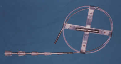
Salem Stripper
<< Back to Operation introdunction || Continue Operation Technique >>
|